Paediatric melanoma
Introduction to the human skin
In order to understand what paediatric melanoma is, it is important to understand where and how it does originate.
Brief overview
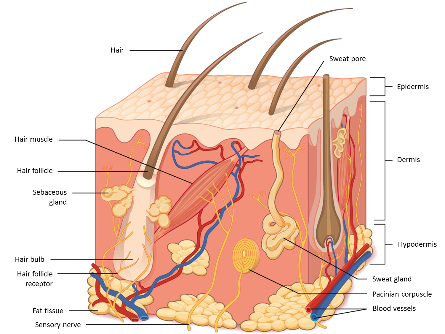
The skin constitutes 16%1 of the human body weight and is one of the tissues in our body that duplicates the most and the fastest. Skin is formed by three main layers:
- The epidermis: Its main job is to protect the body and help control body temperature. It is made up of four types of cells, including pigment cells called melanocytes (see next page).
- The dermis is much thicker than the epidermis. It contains hair roots, blood vessels, lymph vessels, glands, and nerve endings. Blood and lymph vessels in the dermis bring nutrients to the dermis and epidermis. Glands make fluids the body needs, and the connective tissue (which builds an extracellular matrix) holds all these structures in place and allows the skin to stretch.
- Hypodermis is the subcutaneous tissue, what means “below the skin”. It is mostly made of fat and connective tissue. It connects the skin to muscles and bones and it also saves body heat, stores energy, and absorbs shock to protect the body from injury.
The dermis and adjacent fatty tissue layers are not visible to the naked eye. Skin is rich in cell types that have the potential to grow cancer if exposed to repeated ultraviolet trauma, such as excessive sun exposure.
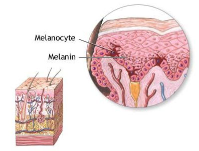
Melanocytes are located at the bottom of the epidermis. These cells make a pigment called melanin, which moves to the top of the epidermis and gives skin its colour. People with darker skin have the same number of melanocytes as people with lighter skin. The darkness of skin is based on how much melanin is made by the melanocytes. Higher levels of melanin cause the skin to be darker.
We lose many skin cells every day, because they complete their life cycle in approximately 20-30 days. The cells that are found in the surface are lost, and cells in the dermis and the basal layer of the epidermis that have a high potential of replication (the ability of the cells to duplicate), replace the cells that are lost in the surface. The cells in the epidermis move from the basal layer to the top (surface), where they are eventually lost renewing constantly the outer layer of our protective skin.
This high potential of replication needs to have a genetic back-up system to minimize the number of errors made in the process of DNA duplication. The genes involved in this mechanism have different functions in sensing and correcting the properly DNA duplication. When these mechanisms don’t work properly, they can give place to diseases such as melanoma.