Xeroderma pigmentosum

Diagnosing skin cancer
In case of suspicious lesions in the skin, to determine if these are cancerous, they are studied via two principal methods: dermatoscopy and biopsy.
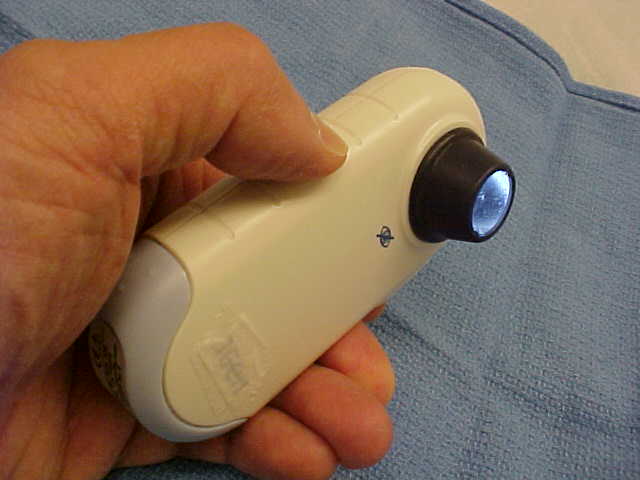
Dermatoscopy is a non-invasive form of examination in which a handheld tool called a dermatoscope is used to assess the skin lesion or mole. It gives an excellent vision of the skin with magnification. The outer layer of our skin is transparent so that the dermatoscope allows the user to assess the patterns of the pigment in the deeper layers of the skin. Thus the experienced examiner is able to see the underlying structures of the lesion.
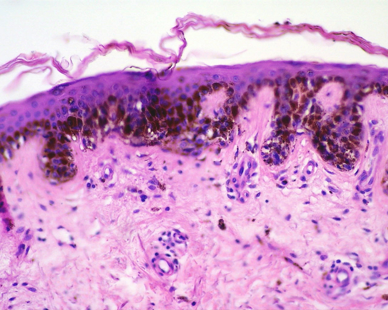
If the lesion seems malignant after dermatoscope, a cutaneous biopsy can be done for further evaluation. This is a form of examination in which a sample of tissue is taken from the suspicious skin lesion and examined under a microscope by a pathologist to determine its abnormal cellular properties.
If during the examination, the sample tests to be malignant, a committee of experts in skin cancer will analyse the case and they will decide together which it is the best procedure to follow.