Retinoblastoma
Introduction to the eye
The eye is a spherical organ located inside the skull (within a bone structure called the “orbit”), which connects to our brain through the optic nerve (ON). We are born with two eyes that allow us to see the world where we live, by decoding a sensorial input (information that enters the eye in form of light) and by “translating” it into electrical information that can travel through the ON to the neurons of our brain. The brain then processes and integrates this data, using it for different purposes, such as helping us see where we are walking, recognize someone’s face, see colours, etc.
Eye’s anatomy and cellular composition
The eye can be compared to an onion, as it is formed by multiple layers on the outside. However, unlike an onion, the inside is full of liquid, which allows the light input to travel through the eye until reaching the internal layer of the posterior wall of the eye, called retina. The retina decodes the light information so it can be eventually processed by the brain.
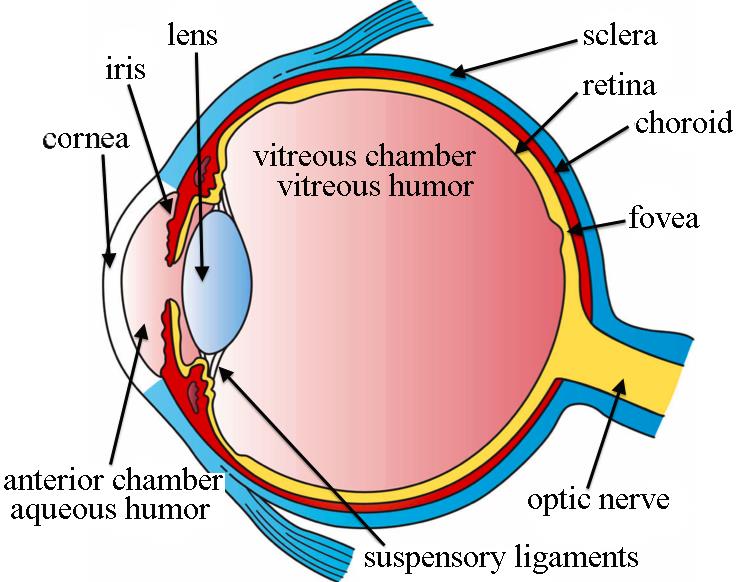
The external layer of the eye is called sclera. Sclera is the white part of the eye that we can see from the outside. The sclera covers the whole eye except the very front, which is covered by the cornea. The cornea is the continuation of the sclera, but instead of being white, it is a transparent layer that allows light to pass through. Together with the sclera, the cornea helps protect the eye against trauma.
The intermediate layer is called choroid, and brings the blood supply to the eye, allowing for the exchange of oxygen and nutrients.
The internal layer, or retina, is actually formed by ten layers of different cells types. The retina is a sensory membrane that covers the inner surface of the eye. The two main types of cells within the retina that function to process light information are called rods and cons. The rods detect motion and allow us to see in black and white when there is not enough light on the outside. The cons, on the other hand, allow us to see colours and help us see better in medium and bright light. The retina is also divided in different areas: the macula with the fovea in the middle and the peripheral retina.
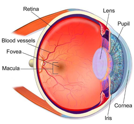
The macula is in the central area of the retina and it provides central vision, which is the high-resolution, colour vision dedicated to the central focus of the eye at any time. In the centre of the macula, there is an area called the fovea, a highly specialized region that provides extreme detail for the input within the eye’s focus. These areas of the retina are mainly formed by cons. The peripheral retina, on the other hand, is formed mostly by rods and provides peripheral vision (what you see around what you are focussing on and what you see in low-light conditions).
The anterior part of the eye is divided into two chambers, both filled with liquid. The anterior chamber is between the cornea and the iris. The lens works just like the lens of a camera, zooming in and out to focus on objects that are closer or further from us. The iris is responsible for the different eye colours. The iris works as the diaphragm of a camera, opening and closing to regulate the amount of light that enters. The posterior chamber is between the iris and the lens. In the posterior part of the eye, the vitreous chamber, between the lens and the retina contains the vitreous humour. The light from the outside travels through the cornea, the anterior and posterior chamber, the lens and the vitreous humour, before reaching the retina, where it will be finally transformed into an electrical impulse and delivered to the brain via the optic nerve.