Osteogenesis imperfecta

2. The bone under the microscope
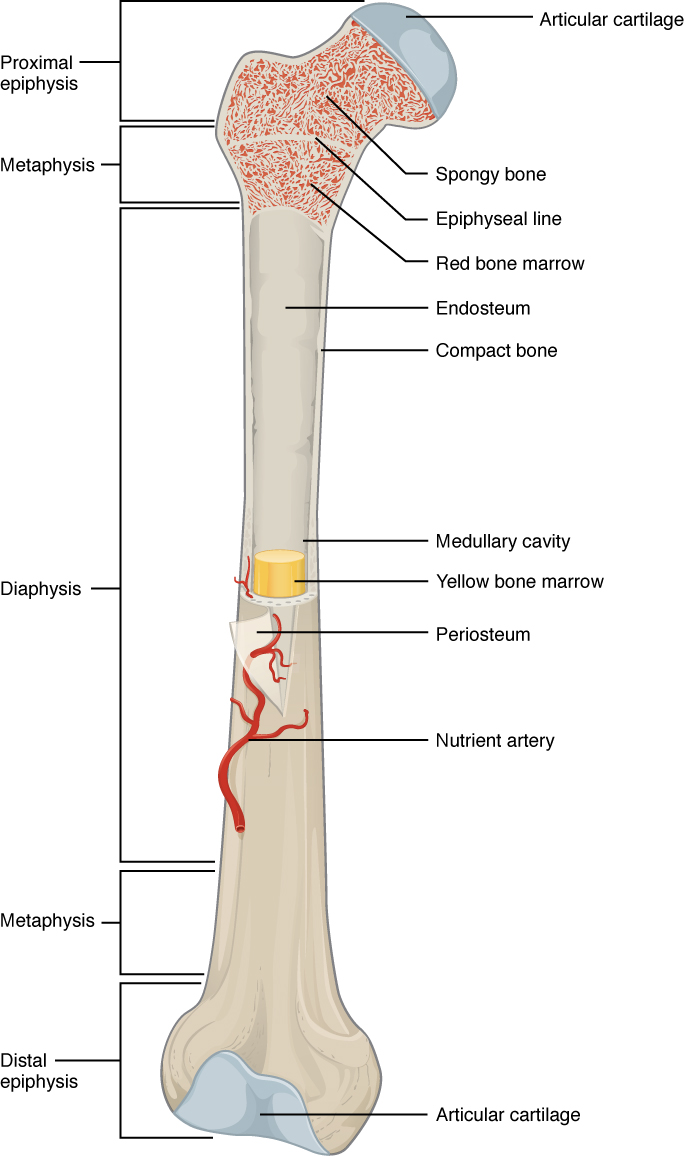
If we cut bone open and look inside, we will see two different textures: the outer part of the bone is the bone cortex which is compact, while the inner part, the trabecular bone, has a spongy texture. The vertebrae are made up mostly of trabecular bone. Inside the bone there is the bone marrow—the tissue responsible for the formation of blood cells, such as the erythrocytes or red blood cells, the platelets, and the leucocytes or white blood cells. The outside of the bone is covered of a thin protective layer called the periosteum (Figure 5).
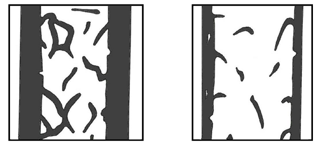
Bones affected by OI usually have a thinner cortex than healthy bones, and the trabeculae are also thinner and smaller in number (Figure 6).
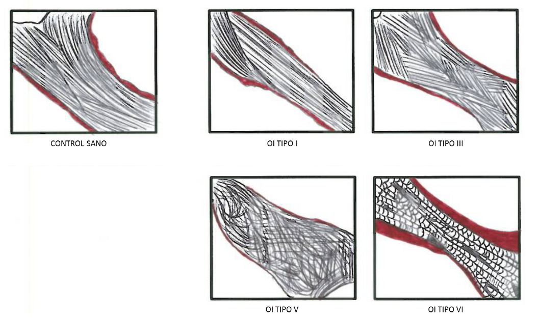
When a fragment of bone is extracted for a biopsy, we can use the maximum magnification of the microscope to see the main cells of the bone – osteocytes, osteoblasts, osteoclasts – which we learned about in the previous chapter. We can also see the collagen fibers arranged in parallel, forming bands called lamellae. Analysis of multiple OI-affected bone biopsies allows to distinguish the histological characteristics typical of certain groups (Figure 7):
- In type I OI, if we observe the collagen fibers under a polarised light microscope, we can see less lamellae but well organised than in bone samples of healthy subjects.
- In type III OI the collagen fibers are somewhat disorganised in their arrangement.
- In type IV OI (mutations in the IFITM5 gene) we see little osteoid tissue (a not mineralised bone matrix) and collagen lamellae are quite disorganised.
- In type VI OI (mutations in the SERPINF1 gene) the bone matrix is under-mineralised, so we see a thick osteoid. The collagen fibers are arranged in a characteristic pattern that reminds of fish scales .