Duchenne muscular dystrophy
Introduction to the muscular system
The muscular system is a set of tissues in the body with the ability to change shape.
The nervous system coordinates the contraction of the muscular system and synchronises functions.
Classification of the muscles
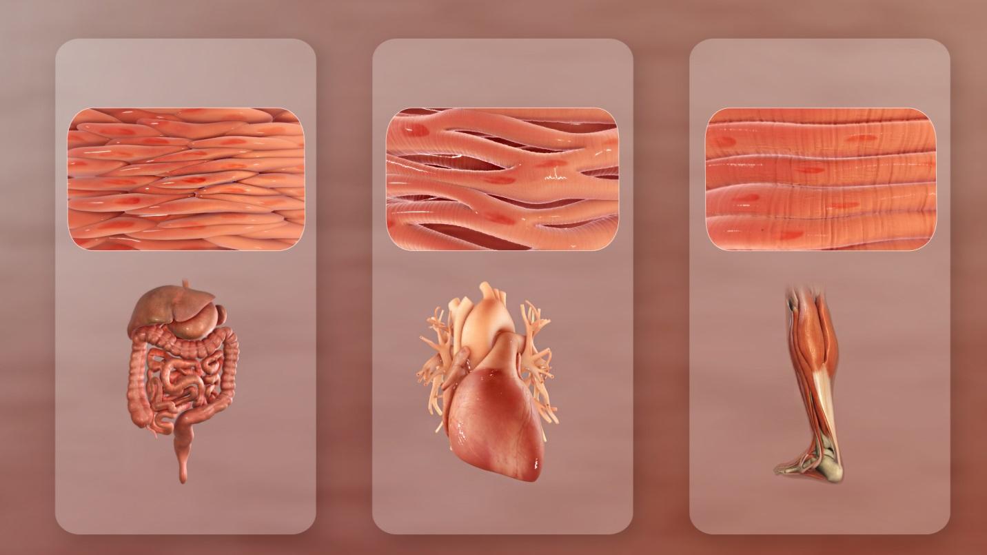
Skeletal or striated muscles attach to the skeleton. They contract to create movement in the body.
The skeletal muscles connect to the somatic nervous system. The somatic nervous system controls voluntary movement of the muscles. Skeletal muscles form part of the musculoskeletal system. Together with tendons and other connective tissues, it connects and secures the joints and the skeleton.
Cardiac muscle makes up the mass of the heart. It causes the rhythmic contractions that pump blood around the body.
Smooth muscles are under autonomic or involuntary control. They exist in the walls of blood vessels. They also exists in structures such as the bladder, the intestines and the stomach. These muscles use less energy, and usually move fluids inside tissues to help shape them.

Cellular composition
Skeletal or striated muscles are responsible for muscle contraction. The main proteins used are actin and myosin, which connect various parts of the muscle cells.
When these proteins receive energy, they slide past each other. This pulls the ends of each muscle cell together. The sarcomeres (or function units of actin and myosin) produce the banding seen in striated muscle. The head of the myosin releases the actin. It then reaches forward and reattaches to the actin. This moves the protein filaments and contracts the muscle fibres.
Smooth muscle cells do not contain these stark bands of protein. The actin and myosin fibres work in a different way from skeletal muscle.
What is dystrophin?
Duchenne Muscular Dystrophy (DMD) is the largest known human gene. It contains the instructions for making a protein called dystrophin1. The primary location of dystrophin is in the striated (skeletal and cardiac) muscles. Small amounts of dystrophin are also present in nerve cells in the brain.
In skeletal and cardiac muscles, dystrophin is part of a group of proteins known as a protein complex. They work together to strengthen muscle fibres. Dystrophin protects muscles from injury as they contract and relax.