Osteogenesis imperfecta

1. Eye anatomy and physiology
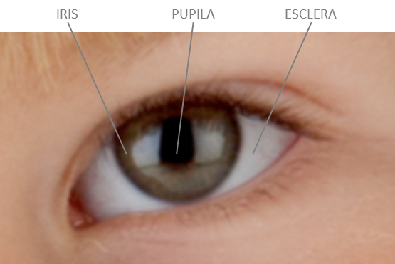
The eye is a complex organ in which the sense of sight resides. If we look at an eye from the front (Figure 1) we can see different parts:
- Iris: this is the part that gives the eyes their colour (green, blue, brown...).
- Pupil: this is a hole in the centre of the iris through which light passes. The iris can control the size of the pupil: when the "hole" is larger, it lets more light through, while when it is smaller, it lets less light through.
- Sclera: the white part of the eye.
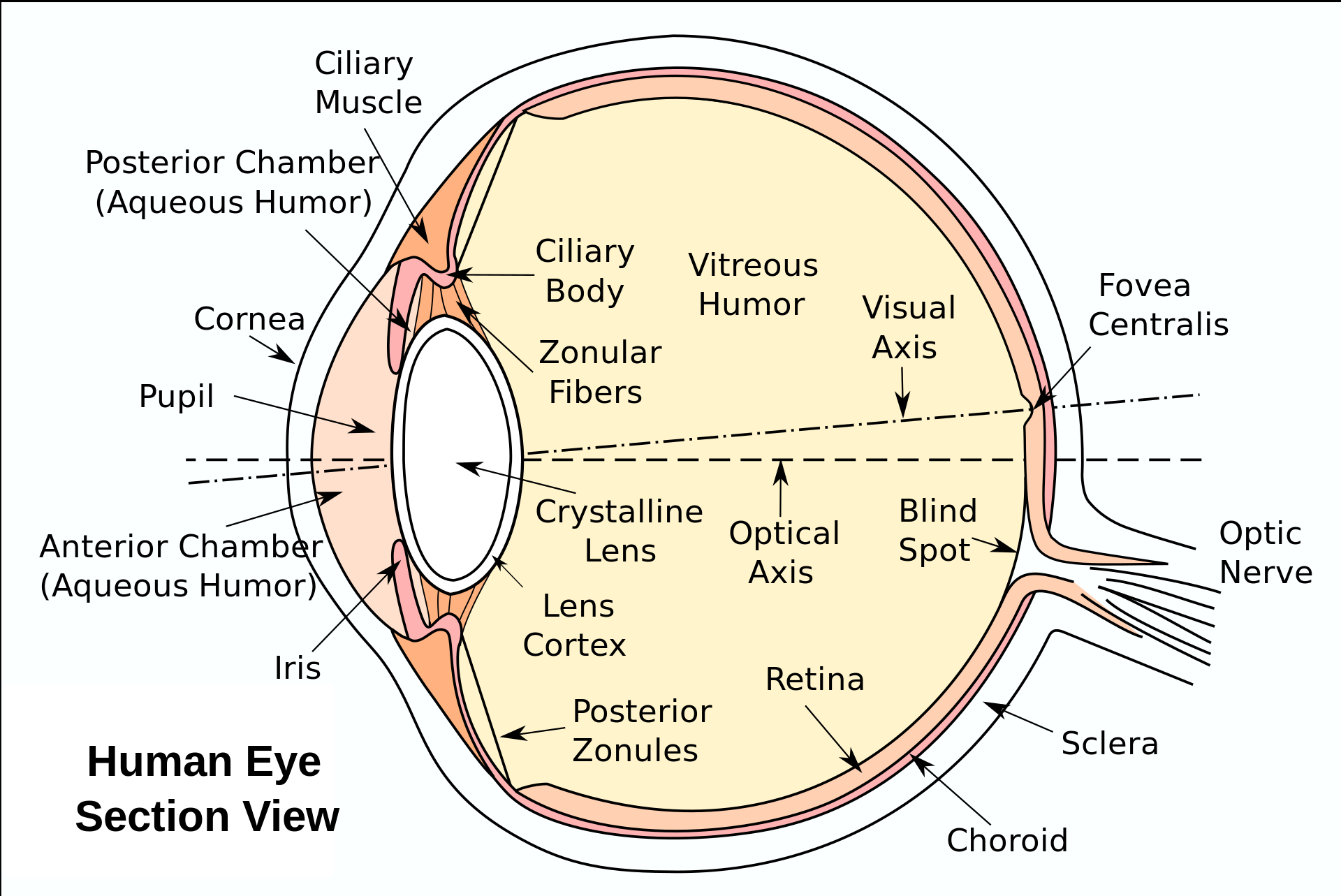
But the eye is really a globe, and if we were to cut it open and look at it in profile we could distinguish the following parts (Figure 2):
How does the eye work?
The eye works in a similar way to a camera.
1. The camera lens
The lens of a camera is a device that contains a set of lenses, which direct the light rays towards the sensor. In the eye, we also have a series of lenses that serve as the lens:
- The cornea is the first lens that encounters the light from outside when it reaches the eye. It is the transparent, domed part at the frontmost part of the eye, and could be compared to a watch glass. It plays an important role in the optical power of the eye. Its transparency depends on a balanced state of hydration. For this, the aqueous humour, in contact with the back of the eye, provides water and nutrients.
- The crystalline lens is the second lens that converges light rays onto the retina (Figure 3). It is located behind the iris and in front of the vitreous. It is attached to the walls of the eye through the ciliary body and ciliary muscle. It has the ability to focus images, depending on whether they are closer or further away (accommodation). With age, this capacity is lost (this is what we know as presbyopia) and it also loses its transparency and is often removed when vision decreases (cataract surgery)
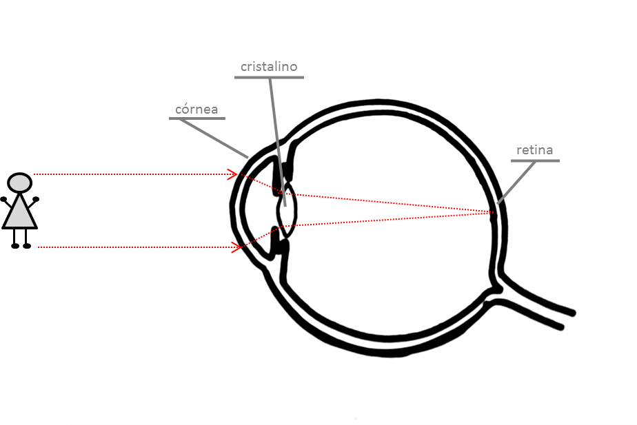
Figure 3. Light rays from the image arrive at the eye, first at the cornea and then the crystalline lens, from where they are projected on to a point of the retina, the fovea centralis, from where they are transmitted to the brain by the optic nerve. Own source.
2. The camera diaphragm
The diaphragm is a device that regulates the amount of light entering the camera by opening and closing it, and also has its equivalent in the human eye.
The iris is a ring-shaped structure that plays the role of a diaphragm, controlling the amount and intensity of light entering the eye. It is also what gives the eyes their colour, and this is determined by the higher (darker eyes) or lower (lighter eyes) melanin (pigment) content, and the arrangement and location of the photoreceptor cells. The circular hole in the centre of the iris is the pupil. Its diameter can vary from 2 to 8 mm depending on light intensity: in bright light it contracts and decreases in diameter (miosis), while in low light or due to the effect of certain medications, it dilates and increases in size (mydriasis).
3. The camera box
The walls of the eyeball are made up of 3 layers:
- The outermost layer, formed by the sclera, which under normal conditions is a thick, tough, white layer.
- The middle layer, called the uvea, includes the iris and the ciliary body in its most anterior part, and the choroid, which is a layer containing abundant blood vessels, in its most posterior part.
- The inner layer is called the retina and is where the light-sensitive cells (the cones and rods) are located.
The eyeball is filled inside by a clear gel called vitreous, which occupies the space between the lens and the back of the eye where the retina is located. In front of the lens there is no vitreous. The anterior chamber is a small space between the lens and the cornea that is bathed in a fluid called aqueous humour. The aqueous humour is formed in the ciliary body, flows into the anterior chamber and is eliminated in a structure located in the angle formed between the iris and the cornea, called the trabecular meshwork. Intraocular pressure is the pressure exerted by the fluids inside the eye to maintain the eye's shape and consistency. It is maintained at optimal and stable values thanks to the balance between the formation and elimination of aqueous humour.
4. Photographic film
The photographic film is the material on which images are processed.
In the eye, it corresponds to the retina, which is the membrane that covers the back of the eye and contains the cells that transform the light received into nerve impulses, which will be transmitted to the brain by the optic nerve. The optic nerve can be compared to an electric cable: it is made up of multiple nerve fibres that originate in the retina and conduct the visual information to the brain, where it is processed (understood, interpreted, stored...).