Hereditary retinal dystrophies

3. X-linked retinoschisis
X-linked juvenile retinoschisis is a vitreoretinal dystrophy characterized by nerve fiber layer fragmentation and intraretinal cyst formation. This disease is inherited through X chromosome, so it affects almost exclusively males, as women can carry a correct copy of the gene in the other chromosome. The described mutation of the RS1 gene encodes retinosquisin, which is expressed only in the retina and is involved in the development and maintenance of retinal architecture. More than 190 gene mutations are known to cause the disease.
Diagnosis is fundamentally based on the clinic. Other tests such as the electroretinogram (ERG) and the optical coherence tomography (OCT) can help in the diagnosis:
- In the ERG, the “negative wave” image appears due to the presence of an “a wave” of normal amplitude followed by a diminished “b wave”.
- The OCT shows the separation of the retinal layers, especially at the level of the nerve fiber layer.
There is currently no curative treatment for the disease and prophylactic treatment of associated complications has not been proven to be effective, so observation and regular monitoring are recommended, as well as treatment of associated complications such as retinal detachment, hemovitreous, or the progression of the retinoschisis towards the macula, when these appear.
Follow-up should be done more frequently, every 4-6 months, in children under 6 years of age, and from that age it could be done annually. We perform refraction controls under cycloplegia, exploration of the anterior segment, eye fundus and macular OCT.
In 2006, a study appeared in which they used 2% dorzolamide in patients with cystic macular lesions and obtained both anatomical improvement in OCT and functional improvement in visual acuity. However, we have not been able to replicate these results in our clinical practice.
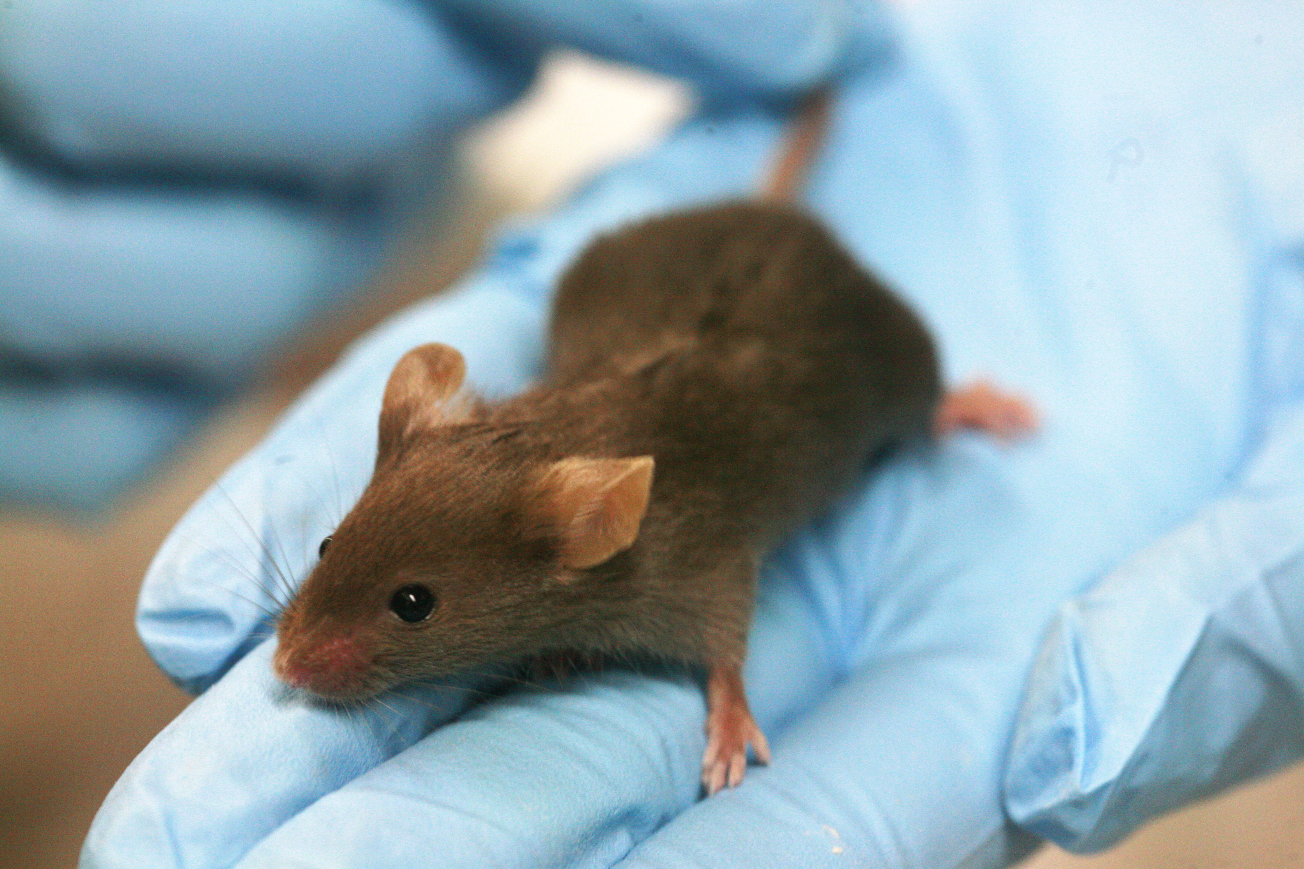
Regarding gene therapy, there are currently 3 animal models for XLRS. The retinoschisin RS1 gene was delivered to affected mice using a viral vector by intraocular injection. Intravitreal administration was shown to be a safe procedure with minimal intraocular inflammatory reaction. It was then shown that retinoschisin had been expressed in the retinal layers and a recovery of the b wave was seen in the ERG. Other works did not obtain such good results with the same procedure.
There are some limitations in the animal model, such as the fact that mice do not have fovea, so we cannot translate the results into what we hope to obtain in humans, since in many patients the disease is restricted to the fovea and, additionally, the mice mutated do not express the retinoschisin protein, unlike humans who do express although it is not functional, so that we do not know if the presence of this abnormal might interfere with the retinoschisin produced by gene therapy.
Even so, given the good results obtained in the tests conducted in animal models, new clinical trials in humans have been initiated.