Hereditary retinal dystrophies

1. The biology of vision
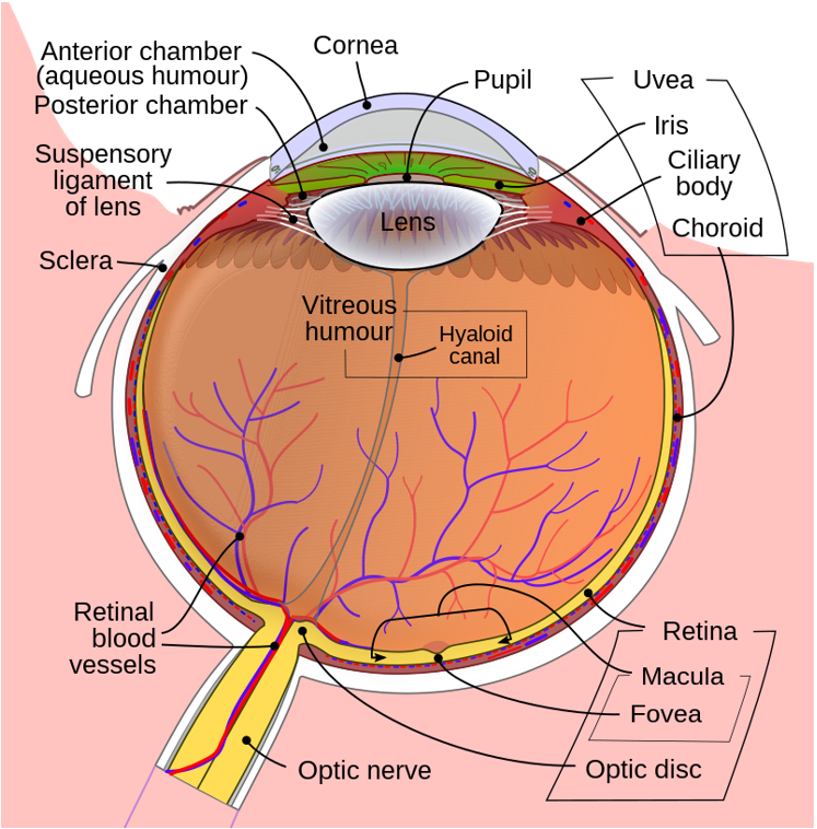
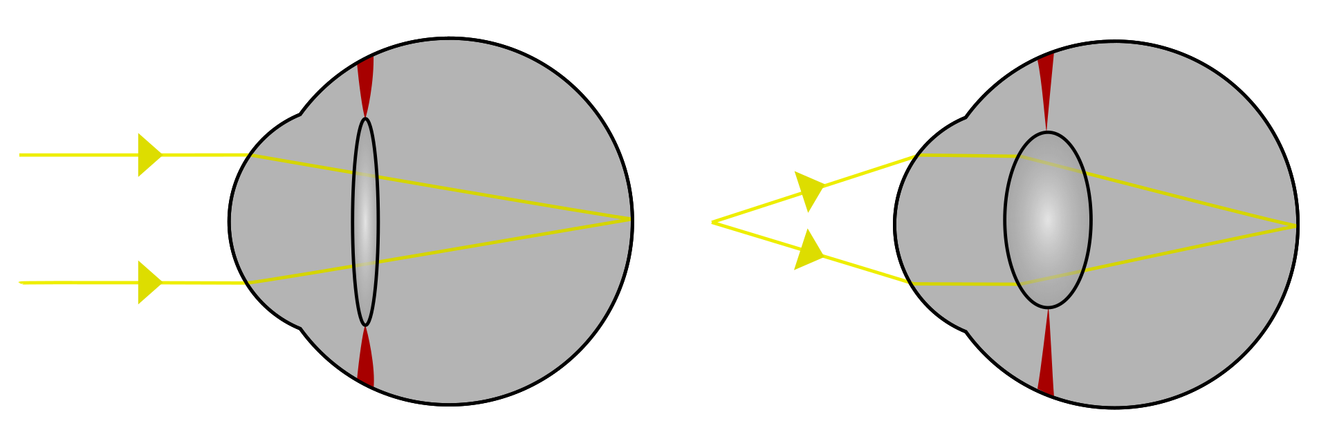
The eye is the organ responsible for vision and it can be compared to a photographic camera. Visual stimuli pass through its transparent parts (cornea, crystalline) before stimulating the visual cells located in the retina, which can be in turn compared to a photographic film. These visual stimuli are then transported through the optic nerve towards the brain, which is in charge of deciphering an analyzing this information.
The visual field stems from these visual stimuli. The amount of light that enters the eye is regulated through the pupillary orifice located in the center of the iris (the colored part of the eye), which works like the diaphragm of a photographic camera, contracting and dilating this orifice.
In the following pages we analyze each of the structures of the eye following a scheme from the outside to the inside of the eye.
The "lens"
Cornea
The cornea is the first “lens” that encounters the light coming from the outside. It is the transparent and convex part located in the most anterior part of the eye. It can be compared to the glass of a wristwatch.
It plays an important role in the optical power of the eye. Its transparency depends on a balanced state of hydration. The aqueous humor, in contact with the posterior portion, plays an important role in providing water and nutrients.
Crystalline
The crystalline is the second “lens” that makes light rays converge upon the retina. It is located behind the iris and in front of the vitreous. It is attached to the walls of the eye through the ciliary body and the zonule, and it has the capacity to focus images (accommodation), an ability that is lost with age (presbyopia). It also loses its transparency with age, and it is often removed when vision decreases (cataract surgery).
The "diaphragm"
Iris
The iris is an annular and contractile structure that is part of the uvea. It plays the role of diaphragm and allows for the diameter of the pupil to adapt to the intensity of light. The color of the eye is determined according to the greater or lesser content of melanin (dark pigment).
Mydriasis: dilation of the pupil.
Miosis: constriction of the pupil.
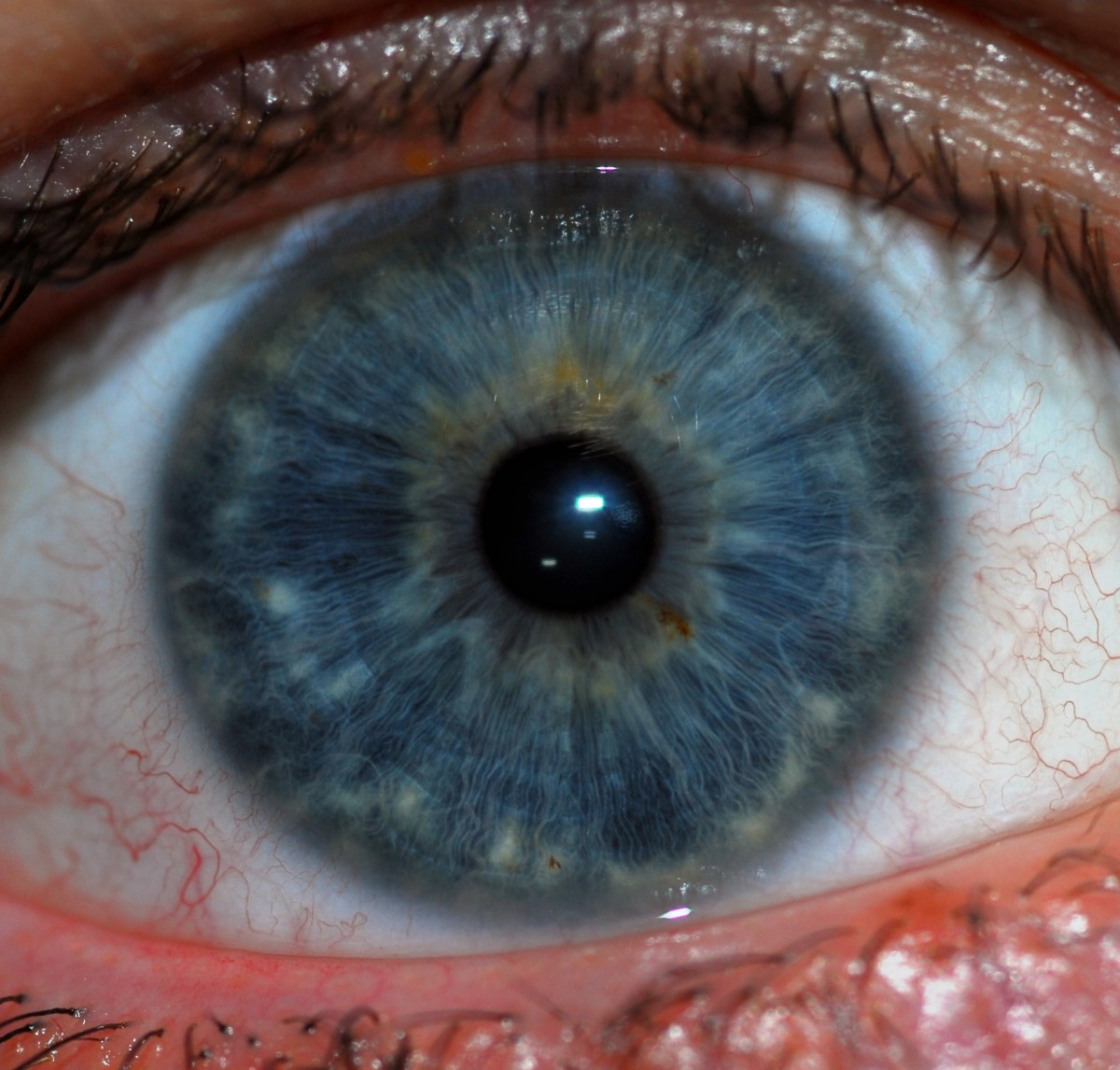
Pupil
The pupil is the circular orifice in the middle of the iris that allows for the light to pass through to the inside of the eyeball. Its diameter can vary from 2 to 8 mm depending on the intensity of light (greater constriction or miosis in situations of much light and greater dilation or mydriasis in situations of darkness), or according to the effects of certain medications.
The "box"
The "box" of the camera is made up of a series of structures and tissues that contain the "lens" and the "diaphragm".
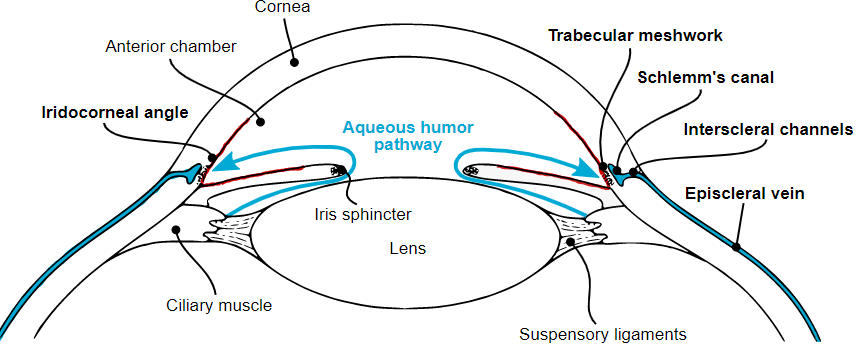
- Iridocorneal angle: the angle formed by the iris and the cornea. At the bottom of this angle is the trabeculum, which is the structure that drains the aqueous humor.
- Anterior chamber: the intraocular space between the cornea and the crystalline, It contains aqueous humor.
- Choroid: the posterior part of the uvea. It is a membrane of vessels and capillaries that play an important role in the nourishment of the retina.
- Ciliary body: a contractile formation that is a part of the uvea where the secretion of the aqueous humor occurs.
- Aqueous humor: a transparent liquid in continuous process of renewal and filtering that occupies the anterior chamber of the eye and bathes both the iris and the crystalline. It is secreted in the ciliary body, and it is reabsorbed in the trabeculum from where it is collected by the Schlemm’s canal and subsequently drained into the venous circulation. The alteration of the balance between secretion and reabsorption causes an increase in intraocular pressure.
- Sclera: the outermost layer of the eye (the white part). White and opaque, it continues forward with the transparent cornea.
- Trabeculum: a circular filter that allows for the reabsorption of the aqueous humor. It is located at the bottom of the iridocorneal angle. If this filter becomes less permeable or if its functioning is damaged, intraocular pressure rises.
- Uvea: the middle layer of the eye. It is a vascular membrane composed of the iris, the ciliary body and the choroid.
- Vitreous: a transparent gel that occupies the back of the eye, behind the crystalline.
The "film"
-
Macula: the area of the retina located in front of the pupil, responsible for fine vision, near vision, relief vision and color vision.
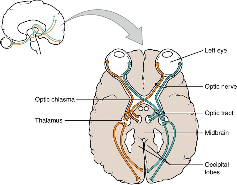
Optic nerve: comparable to an electrical cable, made up by the bundling of visual nerve fibers (about 120,000) originated from the retina It carries visual information towards the brain, where it is processed (understood, interpreted, stored, …) in the posterior region or occipital cortex.
-
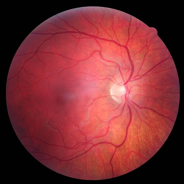
Papilla: the head of the optic nerve visible in the fundus. It corresponds to what is called the blind spot of the visual field since it does not contain “receptors” (retinal cells). It is made up of the nerve fibers that join the retinal cells with the brain.
The physiological cup is a paler area located in the center of the papilla. The fibers arrive from the periphery of the papilla and enter the optic nerve, so that the center can show an “empty” appearance. Normally the size of this cup does not exceed half of the papillary diameter.
- Retina: the membrane that lines the fundus and contains the cells that transform the light received into nerve impulses that will be transmitted to the brain by the optic nerve. It is made up of hundreds of millions of cells capable of capturing light or colours, called photoreceptors.
Photoreceptors
There are two types of photoreceptors: rods and cones. There are more most rods, about 120 million, ans these are more sensitive than the cones. Rods help us see in the dark and are not sensitive to colour.
The 6 to 7 million cones are what give us colour sensitivity and are more concentrated in the macula. In the center of that macula is the so-called fovea, an area about 0.3 mm in diameter, where there are no rods and presents a high concentration of cones.
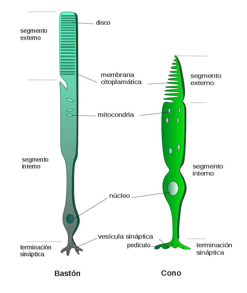
There are three types of cones: those sensitive to red light, those sensitive to blue light, and those sensitive to green light. In the cones we find a sort of flattened sacs that are called membranous discs, where is located the so-called visual pigment capable of modifying its characteristics when stimulated by a certain light (color) and which by a transduction phenomenon gives rise to an electrical signal that will be the one ultimately traveling, together with many other retinal cones signals, along the optic nerve and the optic pathway towards the brain. The brain then interprets colors based on the ratio of stimulation of these three classes of cones.
The rods are the retinal photoreceptor cells responsible for vision at low light levels.
Generally, retinal diseases affecting the cones lead to a loss of central vision and to an alteration in color vision, with frequent photosensitivity (discomfort with excessive bright light levels).
In contrast, diseases predominantly affecting the rods give rise to nyctalopia (low night vision) and to vision loss that begins in the periphery of the visual field.
In retinal dystrophies, we would generally be in the first scenario.
Many of the genes associated to retinal dystrophies are involved in the normal functioning of photoreceptors.
Summary
- The eye is a complex organ that functions like a classical photographic camera.
- At the end of the process, the retina is like the photographic film, being the organ that receives the light and the image.
- The photoreceptors are the retinal cells, highly specialized in transforming the light stimulus into an electrical signal to be carried to the brain.
- Photoreceptors are cones and rods. Cones are capable of giving us information about the color and are concentrated in the central region of the retina. Rods perform better at low light levels (darkness) and are distributed predominantly in the periphery.
- Retinal diseases affecting the cones give rise to loss of central vision and to an alteration of color vision, frequently associating photosensitivity.