Juvenile dermatomyositis

2. Diagnosis. Skin testing tools.
The presence of typical skin lesions establishes the diagnosis of JDM. In all patients with suspected and/or diagnosed JDM, a complete baseline study should be performed to determine the potential involvement of other organs. In case the patient has only muscle weakness or the skin lesions are not typical, a differential diagnosis with other rheumatologic and muscular diseases such as systemic lupus erythematosus, mixed connective tissue disease or other inflammatory myopathies should be performed. Exclusively cutaneous diseases such as atopic dermatitis can produce skin lesions that are sometimes difficult to differentiate.
To help us in the diagnosis, to establish the degree of cutaneous activity and to try to estimate a possible evolution we can use the following tools:
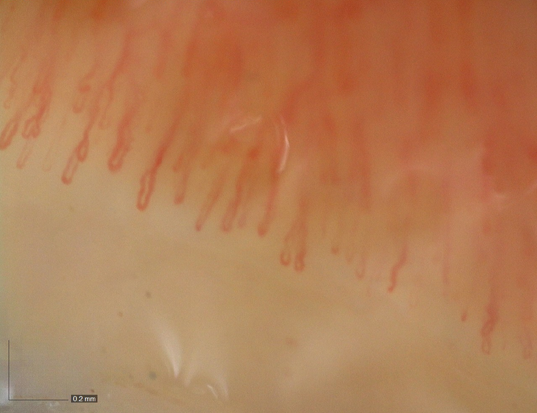
- Capillaroscopy: as previously described, capillaroscopy is a technique that allows us to visualize the capillaries of the nail bed (skin under the nail) with the help of a lens and a light. The inflammation of the capillaries present in JDM is reflected as alteration of their number and/or structure, being able to observe branching, mega capillaries or tortuous capillaries. The alteration of capillaroscopy reflects cutaneous activity although once the disease is controlled, it does not always have to be normalized. There is usually a relationship between the alteration of the capillaroscopy pattern and the severity of the cutaneous disease. There is no specific capillaroscopy pattern for JDM. It is a simple and painless test. To be able to perform it, the nails must be clean, without nail polish application. Your pediatric rheumatologist will apply oil to the nail bed to improve visualization of the capillaries and then assess them through the magnifying lens.
- Myositis-specific antibodies: approximately 70% of patients with childhood inflammatory myopathies have some type of antibody detected, either associated with or specific to the disease. The antibody profile varies according to the disease but the same antibody is usually associated with certain clinical features. Thus, anti-p-155 antibodies are usually associated with severe skin disease, with ulcers, erythroderma, edema and generalized lipodystrophy and anti-NXP2 antibodies with ulcers and calcinosis among other manifestations.
- Skin biopsy: the dermatologist can help us in the diagnosis by performing a skin biopsy. This test consists of extracting a small skin sample and examining it with the aid of a microscope. It is a quick and well-tolerated procedure, as a small anesthetic injection (the only part of the procedure that may be uncomfortable) is given to the area. The skin sample obtained must then be processed for subsequent staining to assess different aspects such as the structure of the skin layers, presence and type of inflammatory cells, presence of vasculitis... Skin biopsy is not strictly necessary for the diagnosis of JDM.
- Measurement tools: the assessment of the extent and severity of cutaneous disease in order to carry out an objective follow-up of the patient remains a problem that has not been fully resolved in JDM. Different measurement tools have been designed, such as the Cutaneous Assessment Tool (CAT) or the Dermatomyositis Skin Severity Index (DSSI), which are usually used exclusively in clinical trials as they are impractical for use in the routine consultation.