Osteogenesis imperfecta

1. Osteogenesis: process of bone formation
Osteogenesis means bone formation. Thus, osteogenesis imperfecta (OI) defines an imperfect bone formation, and it is the term used to refer to a group of genetic disorders characterised in most cases by an alteration in the formation of collagen, a protein that causes bone fragility. This condition is also called “brittle bone disease”.
Bones are part of the skeleton and have several functions:
- Mechanical: bones provide shape and support to the body, enable movement and protect some vital organs such as the brain, heart and lungs.
- Metabolic: the bones are a reserve of ions, mainly calcium and phosphate, which are essential for many activities in our body.
At the microscopic level, the main components of the bone are the cells and the extracellular matrix.
The cells
The main cells of the bone are:
- Osteoblasts: cells derived from stem cells, which synthesise collagen fibres, responsible for the formation of the bone.
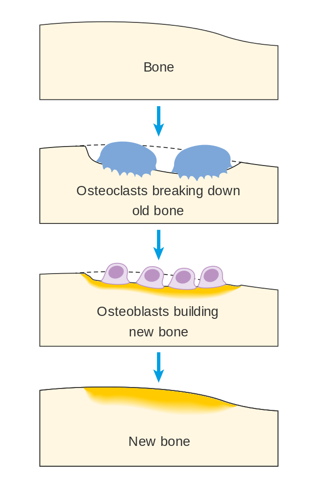
- Osteoclasts: cells that break down the bone tissue.
- Osteocytes: mature cells that do not divide, which derive from osteoblasts and remain “trapped” in the deepest layers of mineralised bone tissue.
Bone is a living and metabolically active tissue that undergoes a constant remodeling process, which gives it a regenerative capacity and functional adaptation. In the process of bone remodelling, osteoclasts located on the bone surface are responsible for destroying the old bone (phase of bone destruction or bone resorption), leaving gaps behind that osteoblasts will later fill with new bone (phase of bone formation), which will then be mineralised (Figure 1).
Through bone remodelling, the bone is able to repair its weathering and meets metabolic needs such as calcium and phosphate release, so that both ions can be used by other tissues. This process is regulated by multiple factors: genetic, exercise, hormones (growth hormone, parathyroid hormone or PTH, and vitamin D…). In order to maintain good bone health, there must be a balance between formation and destruction. In children, the forces of bone formation predominate, while in the elderly the forces of bone destruction predominate.
The extracelular matrix
The bone matrix has two components:
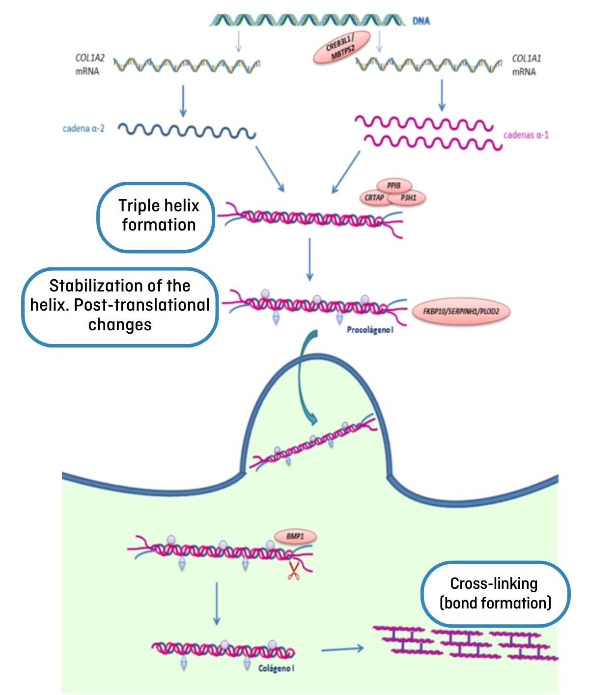
1. Organic: made up of collagen fibres (primarily type I collagen) and non-collagen proteins. Synthesis of collagen begins inside the cell, in a structure called the endoplasmic reticulum. Initially, a pro-collagen I molecule is formed, which is made up of three chains: two identical alpha-1 chains (coded by the COL1A1 gene) and one alpha-2 chain (encoded by the COL1A2 gene), which are assembled forming a triple helix. Multiple proteins act at this level to modify and stabilise the triple helix. The pro-collagen is then transported outside the cell where it is transformed into mature collagen I molecules that gather into fibrils and these in turn are grouped together to form type I collagen fibers (Figure 2). Collagen provides elasticity and flexibility to the bone, preventing fractures.
2. Inorganic: made up of ions and mineral salts that combine to form hydroxyapatite crystals. The main ions involved are calcium, phosphorus, magnesium, fluorine, and potassium. The hydroxyapatite crystals are deposited in the collagen fibers of the osteoid matrix (mineralization process) and the tissue hardens, providing greater strength to the bone.
In most OI cases, there is an alteration of collagen formation — it is either of poorer quality or less than expected — thus, the bone is more fragile and more susceptible to suffer fractures and deformities.